Just
Anatomy
Just Anatomy is just cool
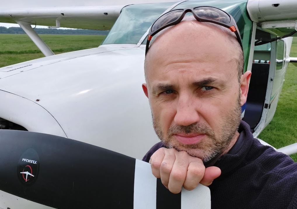
I'm a veterinary anatomist / scientist / researcher whose field of interest collide to ultimately demonstrate the beauty of animal morphology. You could say I have my head in the clouds! Indeed, officially it is called anatomy but this is far too earth-bound an explanation for what I do.
I have a background as Doctors of Veterinary Medicine and continued down the path of research towards PhD in Veterinary and Animal Sciences. I worked together with dr. Kalman Czeibert (DVM, PhD) in several projects (some related images can be seen in the photo gallery), and I have a Veterinary Anatomy teaching experience in both University and Private School settings.
My prime focus is on Veterinary Research where one could integrate and combine the traditional anatomical knowledge with state of art 3D reconstruction and modeling using data from a variety of diagnostic modalities such as CT, MR, surface scanning and cryosectioning. Additionally, I do teaching (either on- or offline).
I create anatomical specimens using traditional skeleton preparations, corrosion casting technique and glass clear resin embedding.
About
Örs Petneházy
I graduated as a vet in 1998. Afterwards, I've worked at the Anatomy and Histology Department of the Veterinary University in Budapest as an anatomy teacher. I have been involved teaching of anatomy and topographic anatomy of domestic animals and birds for Hungarian, German and English student. In 2005 I joined a research group for preclinical human large animal studies at the Diagnostic, Imaging and Research Center of the Kaposvar University. I was involved in different research activities including AMI (artificial myocardial infarct), stent implantation, artificial valve development, CT and MR based large animal research activities. . I defended my PhD in 2012 in cardiovascular analysis of the domestic turkey. Between 2014-2015 I was an assistant professor for veterinary anatomy and diagnostic imaging at the University Alaska Fairbanks (UAF). In 2015 I continued my work at the research group again at Kaposvar.

People
My primary goal in the veterinary anatomical education is to make the complex anatomical stuructures easily understandable by:
- assisted hands on practical dissections on embalmed cadavers (looks very natural but without the annoying smells of a fresh material or formaline)
- high quality video tutorials
- cross sectional images, CT and MR images in multiple planes
- 3D reconstructions and models.
Education
Online or in person courses (practical, assisted hands-on dissection) with detailed explanations on interactive high quality video tutorials for groups or individuals.
Online courses or in person (practical, hands-on dissection and training) courses for veterinarians in various field of the veterinary anatomy: surgical anatomy, cross-sectional anatomy for the CT and MR radiology, X-ray anatomy etc.
3D modeling of various anatomical structures and 3D printing.
Preparation of anatomical specimens:
- High quality skeletal material: complete skeletons, skulls, separate bones. All our bones are professionally macerated, degreased and whitened. All bones are ethically sourced.
- Vascular (corrosion) casts of complete body regions or organs
- Cross sectional atlases
- 3D modeling and 3D printing of various complex anatomical regions

